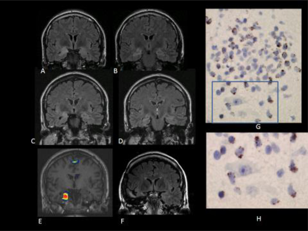
Figure 1.
A, B: Coronal FLAIR MRI images showing increased signal and volumen of right amígdala and hippocampus, suggestive of limbic encephalitis, in a patient with acute onset of seizures. The patient had anti Ma2 antibodies and a past history of seminoma (patient 1, see table).
C, D: Coronal FLAIR images one year later, showing atrophy of right mesial temporal structures with persistence of the increased signal (right MTS) (patient 1)
E: SISCOM showing increased ictal perfusion over the right hippocampus and parahippocampal gyrus during a right temporal lobe seizure with epigastric aura, piloerection and loss of awareness (patient 1).
F: Coronal FLAIR image showing resection of the temporal pole and the right mesial temporal structures. The patient continued to have seizures after surgery although with some seizure reduction (Engel´s class III) (patient 1)
G: Inflammatory infiltrate in the surgical specimen. The tissue section was immunostained with TIA-1 antibody, a marker of cytotoxic T cells. There are positive cells clearly showing the characteristic TIA-1 granular pattern in the inflammatory infiltrate. Some TIA-1 positive cells are in close apposition with neurons (H). (patient 1)

