Figure 3.
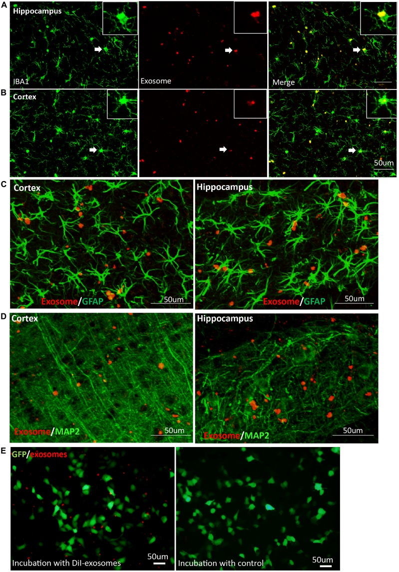
Exosomes target microglia preferentially and stably. Images were captured at 20 days after DiI-exosome injection in non-transgenic mice (17 months old). (A,B) Microglia in the hippocampus and cortex were stained with anti-Iba1 antibody. Merged figures show that the majority of exosomes are localized in microglia. Inset, higher magnification of an exosome-containing microglia cell labeled with Iba-1 antibody. (C,D) Exosomes (red) are not taken up by astrocytes labeled with anti- glial fibrillary acidic protein (GFAP) antibody (green, C) or neurons labeled with anti-MAP2 (green, D) in cortex and hippocampus. (E) SH-SY5Y cells were incubated with DiI-exosomes in vitro. After 24 h incubation, images were acquired using an inverted microscope. Compared with the control group (DiI incubation with PBS and being re-isolated, washed as the DiI-exosomes’ preparation and the same volume of control was incubated with sy5y cells), fluorescent signals are observed in the medium incubated with DiI-exosomes but are rarely detected in SH-SY5Y cells and cannot be detected in the control group.

