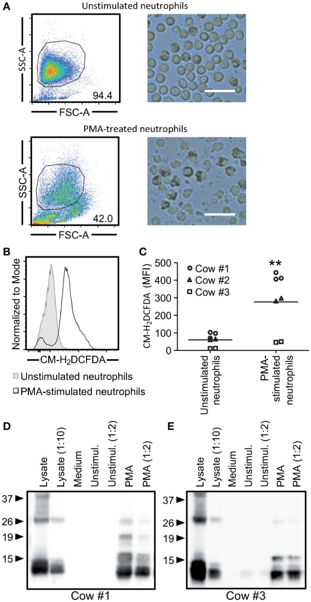
Figure 6.

Cathelicidins can be detected in the supernatants of phorbol myristate acetate (PMA)-activated neutrophils. A total of 5 × 10⁷ neutrophils were stimulated with 100 ng/ml of PMA for 10 min, washed, and cultured for 1 h followed by analysis of reactive oxygen species (ROS) and cathelicidins. (A) Forward and side scatter characteristics of neutrophils stimulated with PMA or left unstimulated were evaluated by flow cytometry (left). Changes in morphology were observed by bright field microscopy with 100 magnification (right). White bars represent 25 µm. (B) Histogram overlay of CM-H2DCFDA fluorescence as an indicator of ROS formation. The unfilled histogram represents PMA-stimulated neutrophils and the gray-filled the unstimulated cells. Cow #1 is shown. (C) The graph shows mean fluorescence intensity of three independent animals each performed in duplicates. Each symbol represents an animal, and the line shows the average of all animals. Significant difference in the average was observed using a paired t-test (**p < 0.001). (D,E) PMA-stimulated neutrophils released cathelicidins into the medium. Undiluted and twofold diluted neutrophil supernatants were employed for western blots using a rabbit anti-bovine cathelin antibody (10 µg/ml). Cow #1 and cow #3 are shown on the left and right side, respectively. Neutrophil lysates were used as positive controls.
