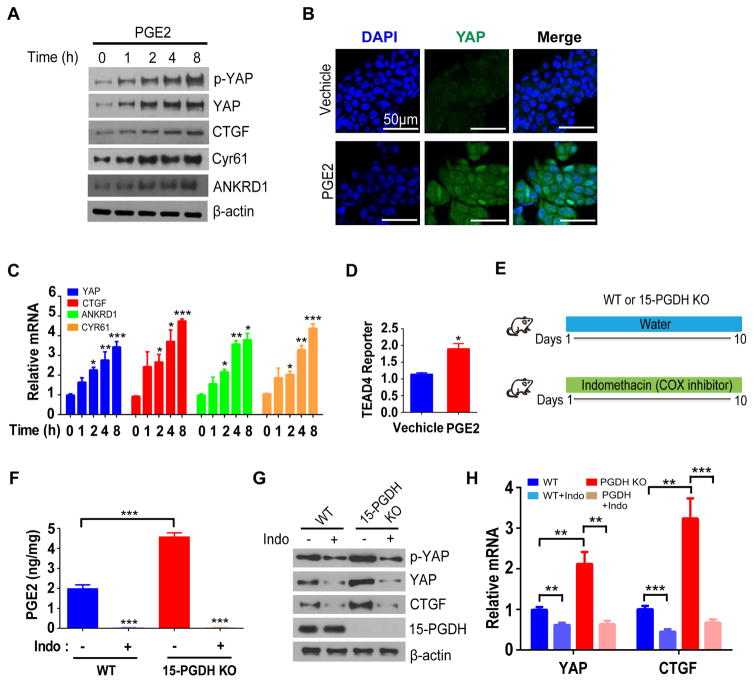
Figure 1. PGE2 positively regulates YAP expression in human colon cancer cells and mouse colon.
(A) Immunoblot analysis of DLD-1 cells that had been deprived of serum for 24 h and then exposed to PGE2 (10 μM) for the indicated times.
(B) Immunofluorescence staining of YAP in serum-deprived DLD-1 cells exposed to PGE2 (+) or vehicle (−) for 18 h. Nuclei were stained with 4,6-diamidino-2-phenylindole (DAPI). Scale bars, 50 μm.
(C) Reverse transcription (RT) and quantitative polymerase chain reaction (qPCR) analyses of samples from A.
(D) Relative TEAD4 transactivation activity in DLD-1 cells transfected with a luciferase reporter construct containing eight tandem TEAD binding sites, deprived of serum, and then exposed to PGE2 (+) or vehicle (−) for 24h.
(E) Experimental protocol for manipulation of PGE2 levels in the mouse colon by 15-PGDH knockout and indomethacin (Indo) administration.
(F) PGE2 content as determined by mass spectrometry in the colon of WT, WT + Indo, 15-PGDH-KO, and 15-PGDH-KO + Indo mice.
(G, H) Immunoblot (G) and RT-qPCR (H) analyses of the colonic mucosal epithelium and crypts of the indicated groups.
All quantitative data are means ± s.e.m. (n = 5, 7, 5, and 5 in C, D, F and H respectively). *P< 0.05, **P< 0.01, ***p< 0.001 versus the indicated comparisons (two-tailed Student’s t test).

