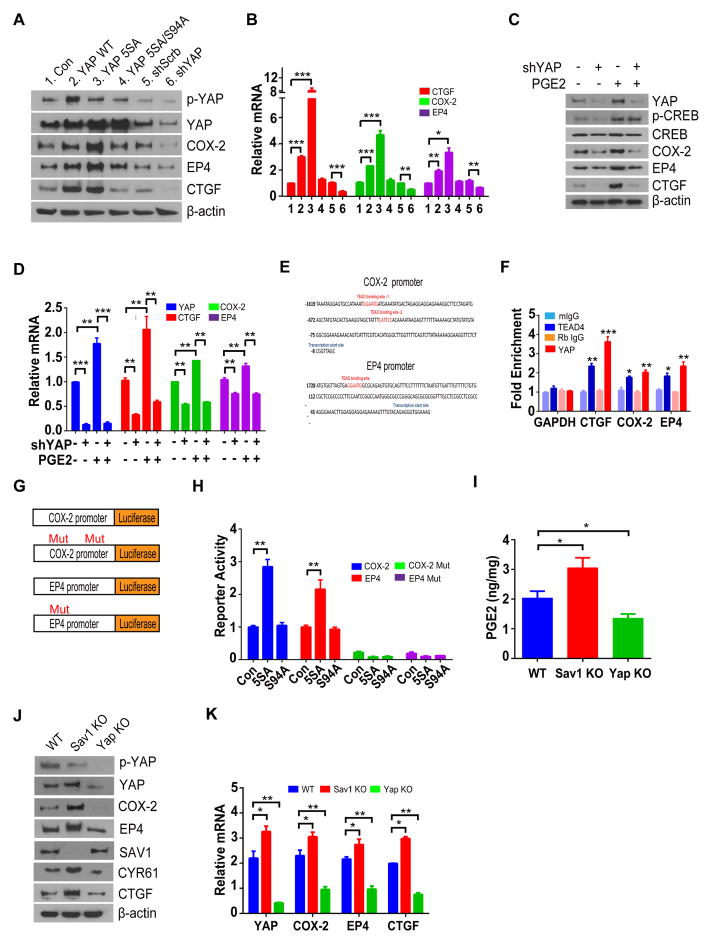
Figure 3. YAP reinforces PGE2 signaling by forming a positive feedback loop.
(A, B) Immunoblot (A) and RT-qPCR (B) analyses of DLD-1 cells infected with retroviruses encoding YAP (WT), YAP(5SA), YAP(5SA-S94A), scrambled or shRNA against YAP.
(C, D) Immunoblot (C) and RT-qPCR (D) analyses of DLD-1 cells infected with scrambled or YAP shRNA treated with PGE2.
(E) Putative TEAD binding sites (red) in the promoter regions of human PTGS2 and PTGER4.
(F) ChIP-qPCR analyses of PTGS2 and PTGER4 promoter regions performed with rabbit antibodies to YAP, mouse antibodies to TEAD4, and corresponding control immunoglobulin G (IgG) in DLD-1 cells growing in full-growth medium. Data are expressed as fold enrichment relative to input DNA. CTGF and GAPDH were examined as positive and negative controls, respectively.
(G) Luciferase reporter constructs used.
(H) Luciferase reporter activity was measured in cells expressing indicated plasmids.
(I) PGE2 content of the colon in WT, Sav1-KO, and YAP-KO mice.
(J, K) Immunoblot (J) and RT-qPCR (K) analyses of the colonic mucosal epithelium and crypts of the indicated groups.
All quantitative data are means ± s.e.m. (n = 5 for all experiments). *P < 0.05, **P < 0.01, ***P < 0.001 (two-tailed Student’s t-test) versus the indicated comparisons.

