Fig. 1.
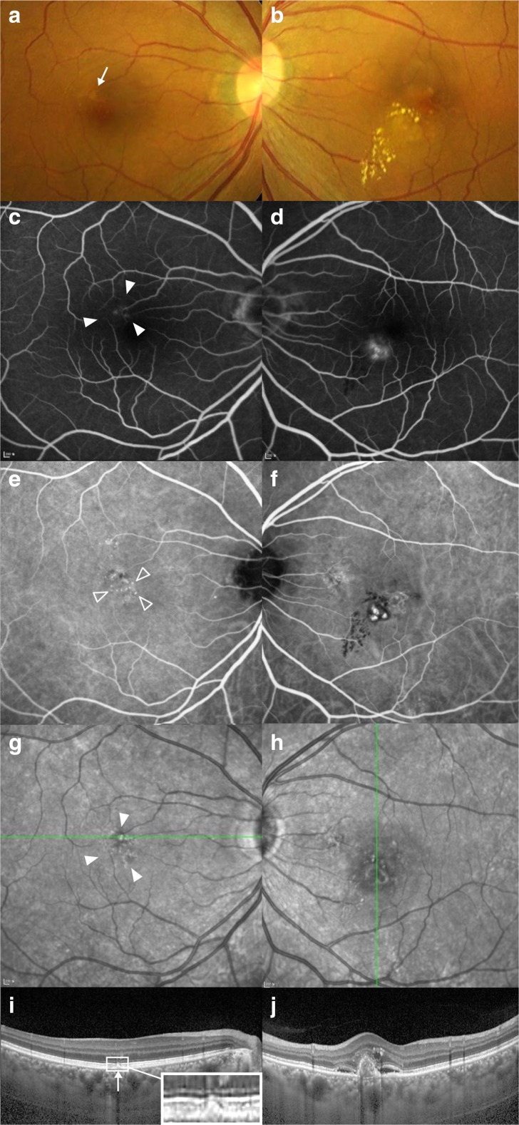
Fundus color photographs (a, b), fluorescein (c, d) and indocyanine green (e, f) angiographs, infrared images (g, h) and optical coherence tomographs (i, j) of a 58-year-old man with unilateral polypoidal choroidal vasculopathy. In the right column, the multimodal images of the left eye with polypoidal choroidal vasculopathy are demonstrated. In the left column, the multimodal images of the uninvolved right eye are demonstrated, which represents the eyes in Group 1. (a) Funduscopic examination reveals drusen-like deposits (DLD, arrow) of a grayish yellow-colored sub-retinal deposit with irregular but discrete margins in the parafoveal area. (c, g) Pigmentary changes (solid arrow heads) adjacent to DLDs visualized by fluorescein angiography and fundus autofluorescence imaging. (e) Mild choroidal hyperpermeability and punctate hyperfluorescent spots (open arrow heads) on indocyanine green angiography. In this case, a DLD is spatially correlated with punctate hyperfluorescent spots (i) Optical coherence tomography scanning over the DLD reveals subretinal deposits, different from that of soft drusen, which usually show dome-like elevation due to sub-retinal-pigment-epithelial accumulation. The subfoveal choroidal thickness is 256 μm

