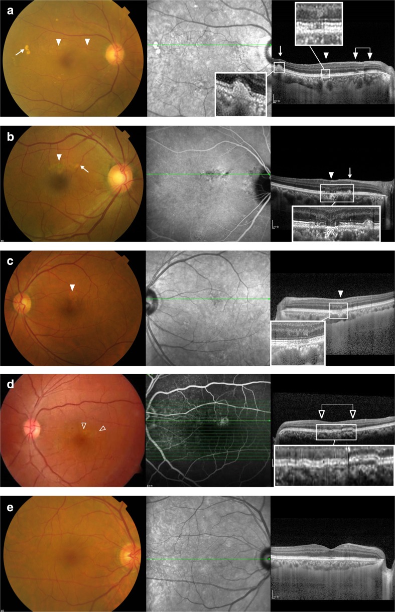
Fig. 2.
Funduscopic and optical coherence tomographic (OCT) images in the unaffected fellow eyes of patients with unilateral polypoidal choroidal vasculopathy are presented. All cases are from Group 1. (a, b) These eyes show drusen-like deposits (DLD, arrows) and pigmentary changes (solid arrow heads). There are DLDs represented by yellowish deposits with irregular but discrete margins. The DLD manifests as an amorphous subretinal deposit usually disrupting the ellipsoid zone on OCT. OCT manifestations of pigmentary changes range from mild attenuation in the interdigitation zone (a) to severe disruption in the outer retina involving the ellipsoid zone and even the external limiting membrane (b). Choroidal thickening is remarkable in both cases. (c, d) These eyes show only pigmentary changes on funduscopy. The third row case exhibits mild disruption in the interdigitation zone and retinal pigment epithelium/Bruch’s complex on OCT. In contrast, a double layer sign (open arrow heads) on OCT is conspicuous in the fourth row case. (e) In a significant proportion of eyes in Group 1, no specific abnormality was noted, as in this case

