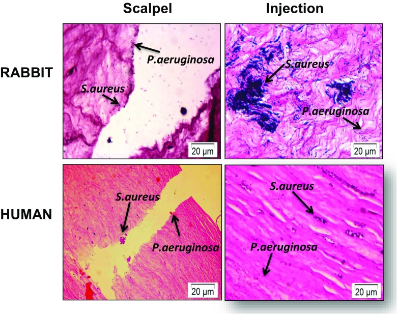
Fig. 6.
Histology of rabbit and human ex vivo models showing a mixed S. aureus and P. aeruginosa infection. At different sites within the same cornea, ex vivo corneas were intrastromally injected with 108 S. aureus and 108 P. aeruginosa and incubated for 24 h (injection). Alternatively, corneas were wounded with a scalpel, and 108 S. aureus and 108 P. aeruginosa were added to the surface of the cornea for 24 h (scalpel). Sections were Gram-stained and imaged to visualise S. aureus and P. aeruginosa within their injection sites at distinct locations within the stroma. P. aeruginosa shows widespread infiltration into the tissue, whereas S. aureus shows less infiltration

