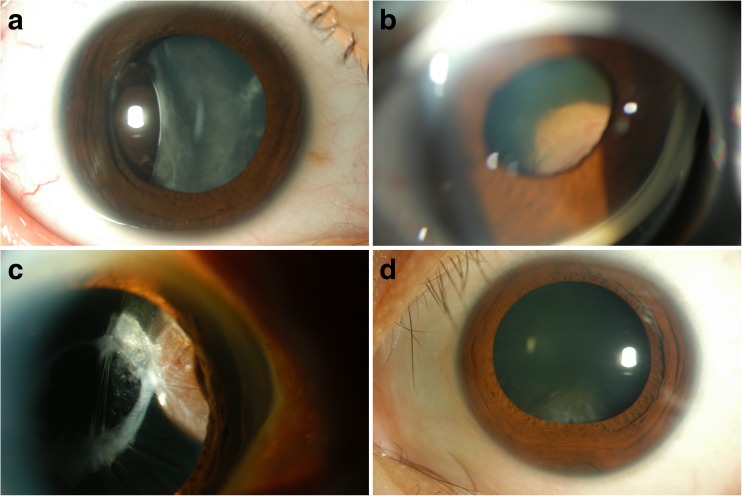
Fig. 1.
Clinical spectrum of benign pigmented intraocular tumors mimicking malignant uveal melanoma. a Case 1: Primary perivascular epithelioid cell tumor (PEComa). Slit-lamp photograph shows a brown mass located temporally, with deformed and tilted lens. b Case 2: Mesectodermal leiomyoma. Gonioscopic photograph shows a mass located in the superior nasal quadrant of the ciliary body, displacing the iris and lens. c Case 3: Melanocytoma. Slit-lamp photograph shows a pigmented mass located in the nasal quadrant of the ciliary body. d Case 4: Melanocytoma. Slit-lamp photograph shows a tumor invading the angle encroaching upon the lens inferiorly

