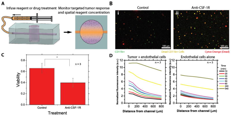
Fig. 3. Treatment with an anti-CSF-1R antibody preferentially causes macrophage death in the DLBCL –on-chip model and diffusion and endothelial permeability can be spatiotemporally modeled.
A) Drug treatments can be infused into the DLBCL-on-chip model and reagent diffusion can be analyzed in conjunction with tumor cell response. B-i) Untreated control devices experience little cell death amongst any DLBCL cell type, B-ii) while anti-CSF-1R antibody treated devices experience preferential cell death of macrophages (CD11b+). Green cells correspond to macrophages (alive or dead), red cells correspond to dead cells of any type and orange cells correspond to dead macrophages. C) Treatment with an anti-CSF-1R antibody causes a nearly 50% reduction in macrophage viability. Statistical significance was determined via t-test (P<0.05). D) Diffusion curves with respect to time and distance of a fluorescently tagged antibody within the lymphoma-on-chip model with and without tumor cells after perfusion for different lengths of time show saturation of the channel within the time scale of the experiment and follow Fick’s law of diffusion. Average normalized fluorescence intensity of 3 experiments is ultimately lower in the case when tumor cells are absent, indicating an increase in endothelial permeability in the presence of tumor cells.

