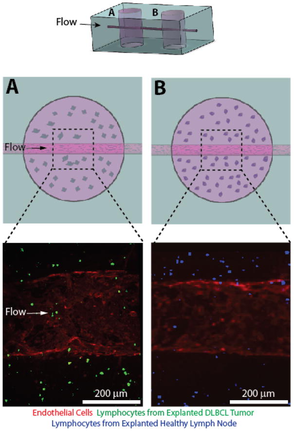
Fig. 5. A multiplex DLBCL-on-a-chip model was developed with a downstream healthy lymph node microvascular model connected via an endothelialized microchannel.
The addition of an additional fabrication step allows the DLBCL tumor hydrogel model (A) to be placed upstream of a healthy lymph node model comprising lymphocytes extracted from an explanted healthy lymph node (B). The top cartoon represents the overall PDMS macrostructure of the multiplex system. The middle row depicts cartoons of the DLBCL tumor hydrogel (A) and the healthy lymph node hydrogel (B). The bottom row depicts confocal micrographs of the distinct microvascular regions illustrated in the row above.

