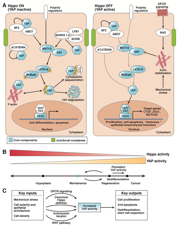
Figure 1. Regulation of the Mammalian Hippo Signaling Pathway.
A. The Hippo pathway (mammalian) consists of the core components STK3 and STK4, SAV, LATS1 and 2, MOB1A and B, YAP, TAZ, and TEAD. Upon activation of the canonical Hippo pathway, STK3/4 phosphorylates and activates Lats1/2, which subsequently phosphorylates cytoplasmic Yap. During homeostasis, Hippo signaling is ON resulting in Yap phosphorylation (S112 in mice, S127 in humans) causing 14-3-3 binding and cytoplasmic sequestration. Phosphorylation of Yap can also lead to proteasomal degradation. When Hippo is off, YAP (in the unphosphorylated for) translocates to the nucleus and binds to the TEAD family of transcription factors, leading to the transcription of genes involved in cell survival, growth, and proliferation. Proposed cell activities for each state are found beneath the gene in italics. Arrows indicate positive relationships, while bars indicate negative activity.
B. YAP activity at various levels and for various time periods differentially modulates cell state and phenotype.
C. Multiple physiologic and pathologic inputs modulate YAP activity. Mechanical stress, cell polarity, and cell density are all factors that have been shown to modulate Yap activity. Additionally, knowledge of these different states is communicated via various signaling modalities, including the aforementioned canonical Hippo pathway, the Wnt pathway, GPCRs, and changes in cytoskeletal tension.

