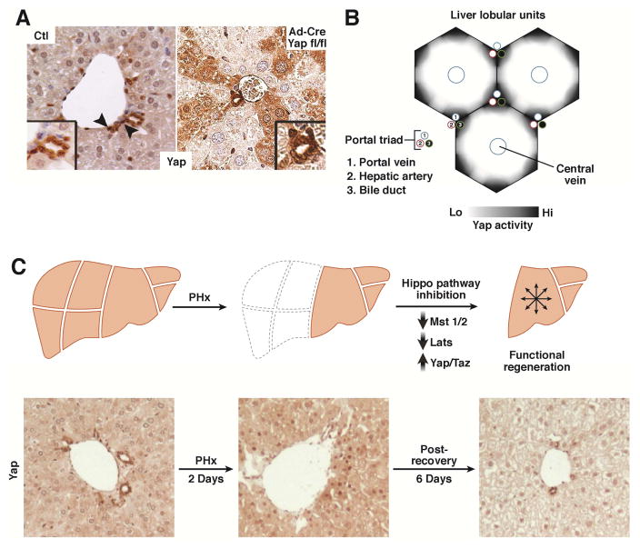
Figure 2. Yap Expression During Homeostasis and Regeneration.
A. YAP is present in the epithelial cells of mouse liver (hepatocytes and biliary cells). YAP expression and nuclear-localization is more prominent in biliary cells (arrowhead) as compared to hepatocytes. Ad-Cre Yap fl/fl illustrates that YAP is present in hepatocytes as documented by mosaic Yap staining after deletion30.
B. Schematic of YAP activity in the liver. YAP activity is highest in the biliary cells/portal hepatocytes, diminishing in the hepatocytes toward the central vein.
C. Hippo/Yap activity dynamically changes after partial hepatectomy. Yap levels increase with an associated decrease in MST1, MST2, LATS1 and LATS2 activity. These return to their normal levels as the liver reaches its appropriate size. Partial hepatectomy in mice results in YAP enrichment and an increase in nuclear localization (Day 2). After 8 days of recovery, YAP expression is reduced to below baseline levels.

