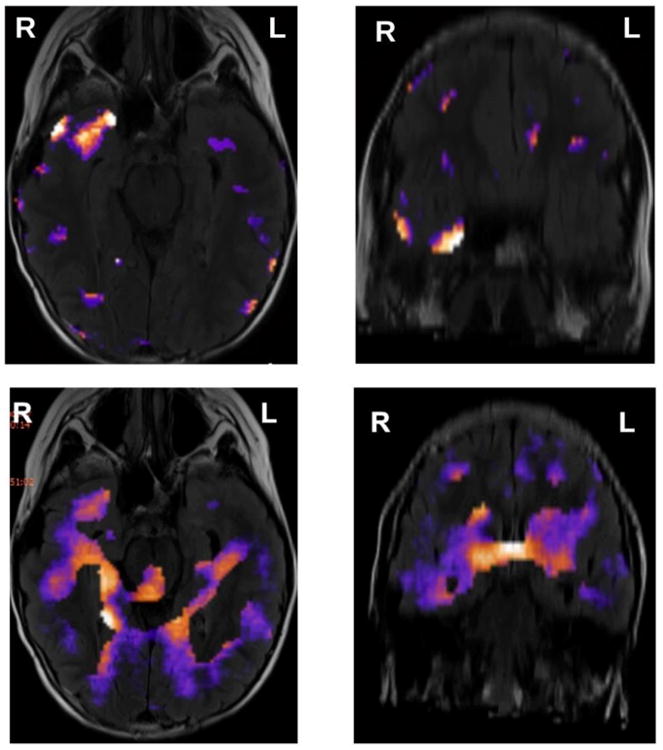
Figure 1.

Examples (axial and coronal views) of (a) successful (focal) SPECT localization and (b) unsuccessful (diffuse) SPECT, from the same pediatric patient with medication intractable focal epilepsy and nonlesional MRI. In the images shown in top panels, the radiotracer was injected 2 s after ictal onset, whereas in the images shown in bottom panels it was injected 30 s after ictal onset, resulting in a focal area of hyperperfusion in the first case and a diffuse area associated with seizure propagation in the second case. The same empirical threshold was used in both cases.
