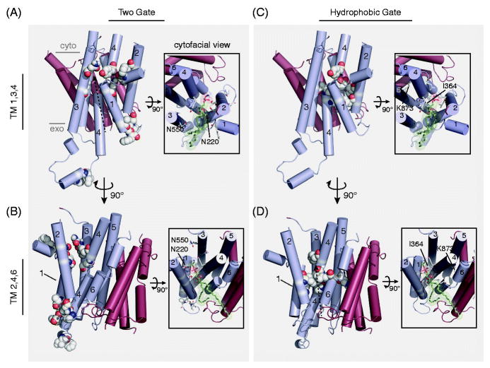
Figure 5. A comparison of putative P4-ATPase substrate pathways.
A,B) The “Two-gate” hypothesis of substrate passage proposes the recognition of PL headgroup at an exofacial/lumenal entry gate (G230, A231, I234, F235, P240, G241, F587, I580) and transport to the cytofacial exit gate (F213, N220, T254, D258, N550, N556, Y618) (numbers from Dnf1). C,D) The “Hydrophobic gate” hypothesis of substrate passage contends that a cluster of hydrophobic residues forming an internal channel to alternate a water-solvated pathway for substrate passage (F88, L112, I115, I362, I364, L367, V908), influenced by the participation of K873 (numbers from ATP8A2). These two hypotheses are not mutually exclusive, and are illustrated by identical dashed-arrows proposing substrate trajectory through the enzyme models. The principle dispute between the two hypotheses is the orientation of the PL substrate during translocation: are the acyl chains directed toward a cleft formed by TM1,3,4 (A,C); or TM2,4,6 (B,D)? Each pathway is depicted using a homology model derived from SERCA (PDB: 3W5D) with a PE molecule docked at the cytofacial aspect in the TM1,3,4 (A,C) and TM2,4,6 (B,D) orientation (Roland and Graham, 2016). Only the TM domain is depicted, with alpha helices shown in cylinders. TM segments 1–6 are colored cyan, while TMs 7–10 are red. Major images: lateral view of the TM domain, substrate-selective residues are colored white/by elements and shown in spheres. Image insets: cytofacial view of the TM domain, substrate-selective residues are colored white/by elements and shown in sticks, docked PE model is colored green/by element and shown in sticks with transparent spheres to illustrate space accommodations. Select residues are numbered and indicated (Dnf1 – Two-gate model; ATP8A2 – hydrophobic gate model). Color figures can be found online.

