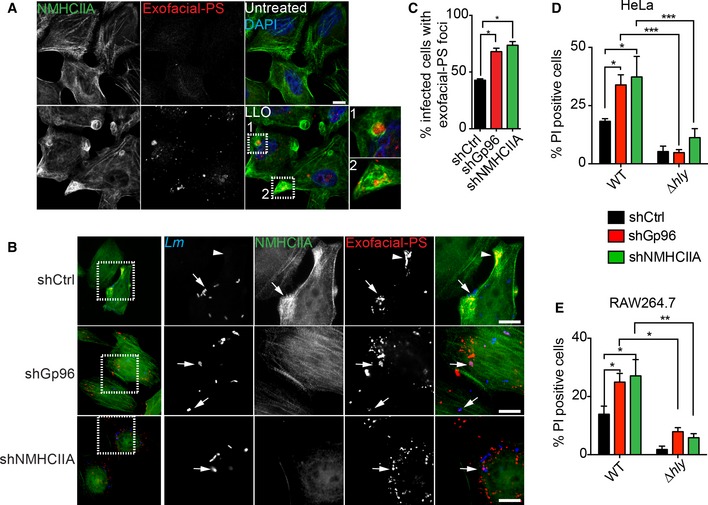
Figure 6. Gp96 and NMHCIIA preserve PM integrity during Listeria monocytogenes infection.

-
A, BEpifluorescence microscopy images of HeLa cells (A) left untreated or treated with LLO (0.5 nM, 15 min) and (B) infected with GFP‐expressing wt Lm for 6 h. Cells were incubated with exofacial‐PS probe (Alexa‐568 annexin A5) (red) 30 min prior to fixation, immunolabelled for NMHCIIA (green) and stained with DAPI (blue). Insets show PM damage marked by exofacial‐PS sites. Arrows indicate exofacial‐PS associated with intracellular bacteria and in shCtrl cells with NMHCIIA bundles; arrowheads indicate NMHCIIA bundles associated with exofacial‐PS sites without detectable bacteria. Scale bar, 10 μm.
-
CQuantification of the percentage of infected cells with exofacial‐PS sites in HeLa cells infected with wt Lm for 6 h. Data are the mean ± SEM (n = 3), and P‐values were calculated using one‐way ANOVA with Tukey's post hoc analyses, *P < 0.5.
-
D, EFlow cytometry analysis of the percentage of PI‐positive shCtrl, shGp96 or shNMHCIIA HeLa (D) and RAW264.7 (E) cells, infected with GFP‐expressing wt or Δhly Lm for 6 h. The levels of PI incorporation by uninfected cells were subtracted from infected samples. Values are the mean ± SEM (n ≥ 4), and P‐values were calculated using one‐way ANOVA with Tukey's post hoc analyses, *P < 0.5, **P < 0.01, ***P < 0.001. PM permeability was assessed in cells harbouring equivalent numbers of intracellular bacteria (Appendix Fig S3).
