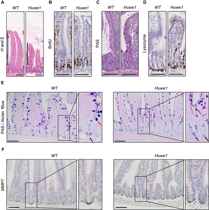
Figure 2. Huwe1 deletion leads to perturbed intestinal homoeostasis.

- H&E staining of control and Huwe1‐deleted intestinal epithelium. Shortened villi in Huwe1‐deficient tissue are indicated.
- BrdU IHC of control and Huwe1‐deleted intestinal epithelium.
- PAS staining identifying goblet cells. No gross changes were observed.
- Lysozyme staining (Paneth cell marker) of control and Huwe1‐deleted small intestine. Note the occurrence of lysozyme‐positive cells away from the crypt base (black arrows).
- Dual periodic acid–Schiff/alcian blue staining to identify Paneth cell secretory vesicles (light blue/pink, marked with black arrows) and goblet cells (dark blue/purple, marked with red arrows). Note Paneth cell secretory vesicles are restricted to crypt base in both control and Huwe1‐deficient small intestines (inset, black arrows).
- MMP7 staining of control and Huwe1‐deleted small intestine. Note MMP7 staining is restricted to crypt base in Huwe1‐deficient intestines.
