
Figure EV6. MCL1 protects Huwe1‐deficient tumours from apoptosis.
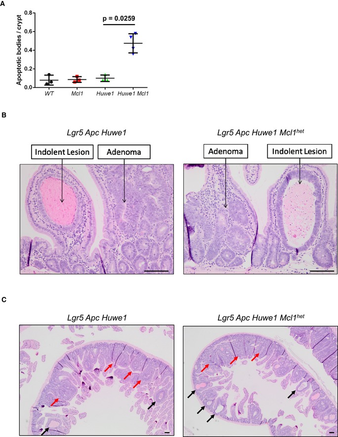
- Quantification of scoring of apoptotic bodies in control, Mcl1fl/+, Huwe1fl/fl and Huwe1fl/fl Mcl1fl/+ deleted intestines (Mann‐Whitney, P = 0.0259, n = 3 vs 4). Data are mean and SD.
- Classification of adenoma and indolent lesions observed in Lgr5 Apc Huwe1 (left panel) and Lgr5 Apc Huwe1 Mcl1het (right panel) mice. Scale bars = 100 μm.
- Image of intestines from Lgr5 Apc Huwe1 (left panel) and Lgr5 Apc Huwe1 Mcl1het (right panel) mice. Adenomas indicated by red arrows and indolent lesions by black arrows. Note the decreased ration of adenomas/indolent lesions observed in Mcl1het intestines. Scale bars = 100 μm.