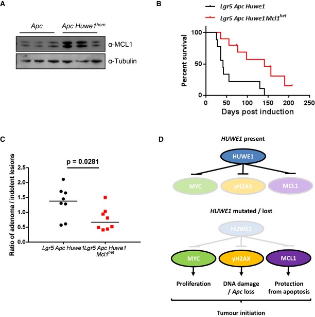
Figure 8. Huwe1‐deficient tumours tolerate high levels of DNA damage due to elevated MCL1.

- MCL1 Western blot in protein extracts from Vil Apc and Vil Apc Huwe1 hom tumours. Levels of MCL1 protein are significantly increased in tumours lacking HUWE1 (Mann–Whitney, P = 0.04, n = 3).
- Kaplan–Meier survival plot of cohorts of induced Lgr5 Apc Huwe1 and Lgr5 Apc Huwe1 Mcl1 het mice. Deletion of one copy of Mcl1 led to a significant increase in survival of these animals (log rank, P = 0.0052, n = 9 versus 10).
- Ratio of adenoma/indolent lesions observed in Lgr5 Apc Huwe1 and Lgr5 Apc Huwe1 Mcl1 het mice. Note the decreased ratio of adenoma/indolent lesions observed upon Mcl1 deletion (Mann–Whitney, P = 0.0281, n = 8 versus 8).
- Schematic outlining the tumour‐suppressive role of HUWE1. When HUWE1 is expressed, the amounts of MYC, γ‐H2AX and MCL1 are maintained at low levels. Following HUWE1 mutation or loss MYC, γ‐H2AX and MCL1 levels are increased leading to increased levels of proliferation and DNA damage. This leads to loss of Apc and tumour initiation. Increased MCL1 is critical to protect these cells from apoptosis induced by the high levels of DNA damage.
Source data are available online for this figure.
