
Figure 6. Analysis of DNA covalently linked with Top1 by immuno‐pull‐down.
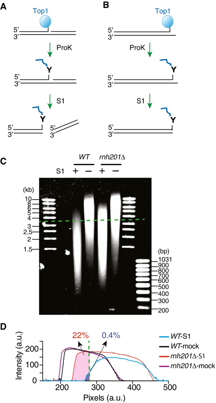
- Diagram depicting the pulled‐down DNA that are covalently linked to Top1 at an internal position. After proteinase K digestion, the resulting DNA nick is cleaved by nuclease S1.
- If the covalently linked Top1 is located at the end and no other nicks are present in the pulled‐down DNA, it is resistant to cleavage by nuclease S1.
- S1‐treated or mock‐treated pulled‐down DNA from either wild‐type (WT) or rnh201Δ strains resolved on a 1% agarose gel and stained with SYBR Gold. A representative gel image is shown, and dashed green line marks the position for DNA of 3.5 kb.
- Traces of DNA intensities for the samples from the gel image in panel (C). Dashed line marks the position for DNA of 3.5 kb, and the listed numbers show the percentage of DNA density longer than 3.5 kb calculated from the total area under the curves.