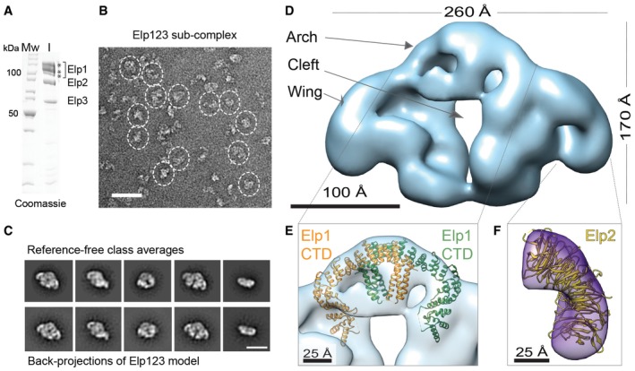
Figure 2. EM reconstruction of endogenous Elp123 sub‐complex.

- SDS–PAGE gel showing the purified Elp123 sub‐complex used as input for gel filtration. Protein bands marked with asterisks presumably correspond to different phosphorylation states of Elp1.
- Representative negative‐stain EM field of the Elp123 sub‐complex. Particles in side and top views are highlighted. Scale bar, 50 nm.
- Reference‐free class averages and back‐projections of the Elp123 model. Scale bar, 20 nm.
- EM reconstruction of the Elp123 sub‐complex at 27 Å resolution.
- Fitting of the Elp1 CTD into the Elp123 reconstruction.
- Fitting of the Elp2 crystal structure in the difference density generated by subtracting the partial Elp123 reconstruction from the complete Elp123 reconstruction.
