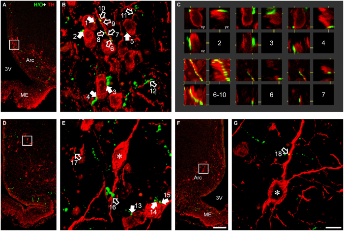
Figure 1. Hypocretin/orexin terminals form close appositions to TIDA neurons.
Low power confocal micrographs (A,D,F) from three different rat Arc sections and levels processed for immunofluorescence for tyrosine hydroxylase (TH; red) and H/O (green). Orthogonal normal shadings of confocal stacks in (B,E,G) represent regions within squares in (A,D,F) respectively. Note close appositions between H/O-immunoreactive (-ir) terminals and TH-ir cell somata (B,E; filled arrows) and dendrites (B, E,G; empty arrows). All close appositions are confirmed in 3D with single optical sections in the xy-, xz- and yz-plane (examples from arrows in B represented in C with corresponding number). 3V, third ventricle; Arc, arcuate nucleus; ME, median eminence. Scale bar in (F) = 100 μm for (A,D,F) and in (G) = 10 μm for (B,E,G).

