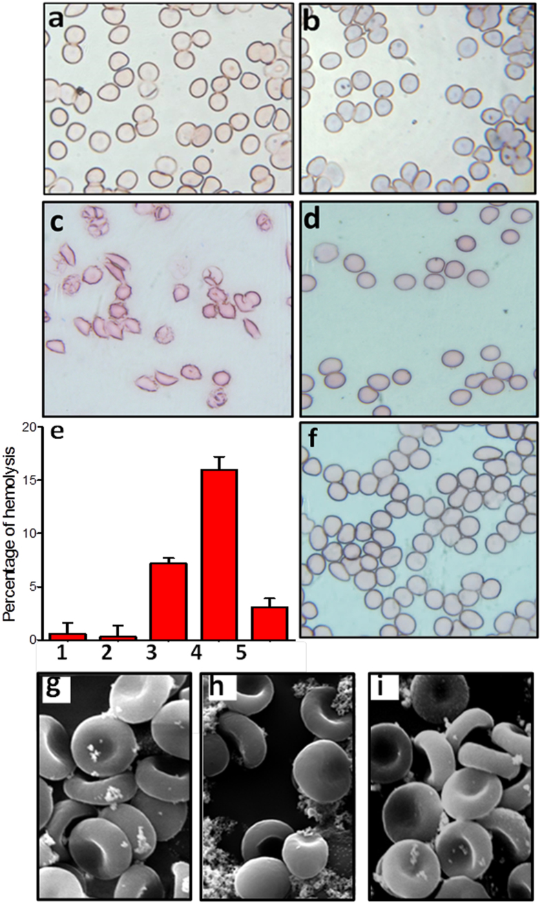
Figure 5. Protective effect of eugenol against amyloid induced hemolysisof RBCs in PBS buffer (pH 7.4) and at 37 °C.
(a) Optical microscopy images of only RBCs in PBS buffer. (b) RBCs + 2 μM of soluble serum albumin. (c) RBCs + 2 μM of amyloid aggregates of serum albumin. (d) RBCs + 2 μM of amyloid aggregates of serum albumin + 0.9 mM eugenol. (e) Histogram showing percentage of hemolysis caused by amyloid fibrils of serum albumin in the presence and in the absence of eugenol: (1) 5 μM soluble BSA; (2) 0.9 mM eugenol; (3) 5 μM aggregated BSA (4) 30 μM aggregated BSA; (5) 30 μM aggregated BSA + 0.9 mM eugenol. (f) RBCs + 0.9 mM eugenol. (g) SEM images of control RBCs. (h) SEM images of lysed RBCs in the presence of BSA aggregates. (i) SEM images of RBCs + amyloid sample in the presence of eugenol.

