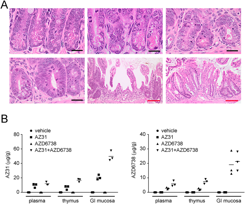
Figure 2. Histopathology associated with AZ31 in moribund mice after TBI and pharmacokinetics of AZ31 and AZD6738 at the time of irradiation.
(A) Photomicrographs of transverse sections of mouse intestine following 9 Gy TBI. Top left: normal small intestinal crypt epithelium following 9 Gy alone (Day 14). Top center: minimal disruption of morphology within the small intestinal crypt epithelium following 75 mg/kg AZD6738 and 9 Gy (Day 14). Top right: disrupted crypt morphology within the small intestine following 100 mg/kg AZ31 and 9 Gy (Day 7). Bottom left: regenerative crypt hyperplasia within the small intestine following 100 mg/kg AZ31 + 75 mg/kg AZ6738 and 9 Gy (Day 6). Bottom center and bottom right: regions of significant atrophy and loss of the crypt epithelium within the small intestine and large intestine, respectively, following AZ31 + AZ6738 and 9 Gy (Day 6). Black bar = 20 μm; red bar = 100 μm. (B) Concentrations of AZ31 and AZD6738 in the plasma, thymus, and small intestinal (GI) mucosa of mice 2 h following a single oral dose of vehicle, 100 mg/kg AZ31, 75 mg/kg AZD6738, or 100 mg/kg AZ31 + 75 mg/kg AZD6738. N = 3 mice per treatment group.

