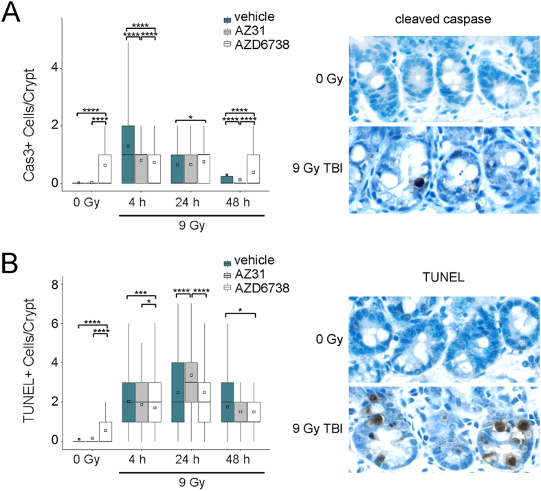
Figure 4. Impact of ATM and ATR kinase inhibition on intestinal crypt cell death after TBI.
Mice received vehicle, 100 mg/kg AZ31, or 75 mg/kg AZD6738 2 h prior to 9 Gy. Small intestine tissues were harvested at the specified timepoints after TBI. For un-irradiated control mice, tissues were harvested at 6 h after inhibitor dosing, equivalent to 4 h after TBI. (A) Left: Enumeration of the number of cleaved caspase (Cas3) positive cells per small intestine crypt in vehicle, AZ31, or AZD6738 treated mice at 4 h, 24 h, and 48 h after 9 Gy TBI, compared to 0 Gy controls. Right: Representative images of cleaved caspase positive IHC staining in the small intestine crypts of vehicle treated mice at 24 h after 9 Gy TBI, compared to 0 Gy control. (B) Left: Enumeration of the number of TUNEL positive cells per small intestine crypt in vehicle, AZ31, or AZD6738 treated mice at 4 h, 24 h, and 48 h after 9 Gy TBI, compared to 0 Gy controls. Right: Representative images of TUNEL positive IHC staining in the small intestine crypts of vehicle treated mice at 24 h after 9 Gy TBI, compared to 0 Gy control. For Cas3 and TUNEL quantitation, box and whisker plots depict counts from a total of 200 crypts (n = 200), with 100 crypts from each of 2 mice. *p < 0.05, ***p < 0.001, ****p < 0.0001.

