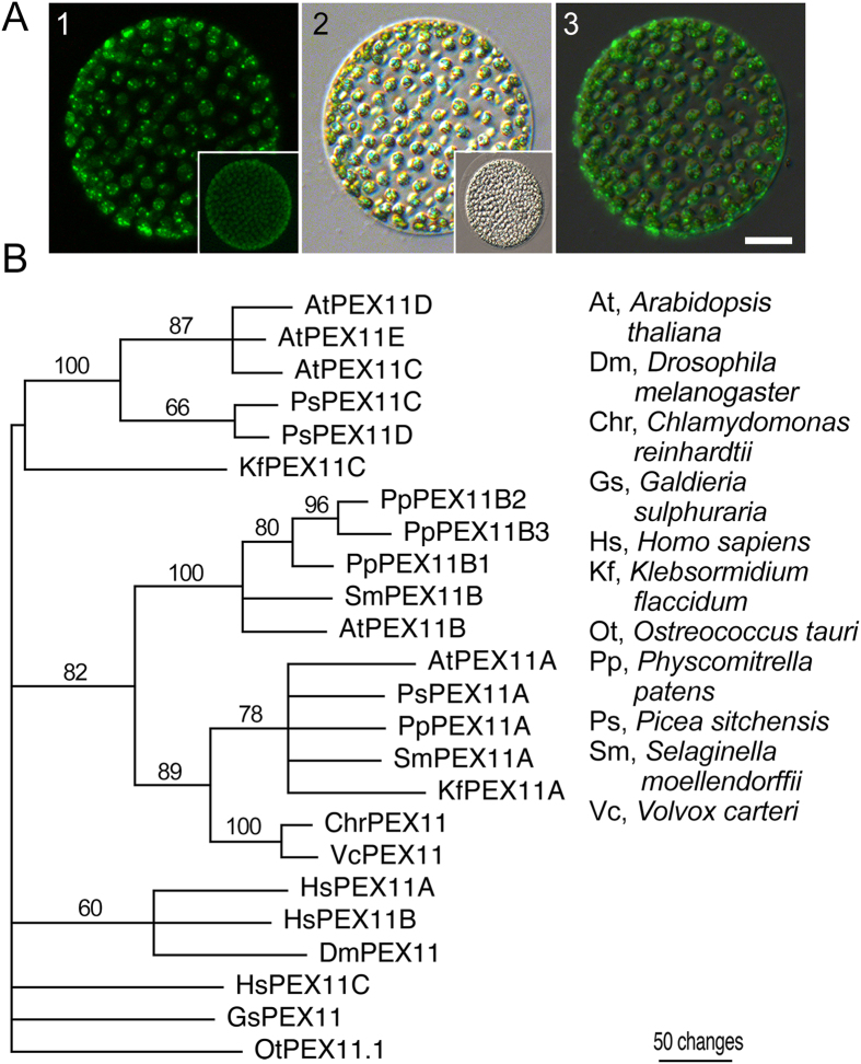
Figure 5. N-BODIPY staining of Volvox sp.
(A) Fluorescence (1), bright field (2), and merged (3) images of Volvox sp. Scale bar 50 μm. Insets show unstained coenobium imaged at the same gain and exposure settings as the main images. (B) Phylodendrogram of a peroxisome biogenesis protein PEX11 from divergent lineages. A PEX11 homologue from Ostreococcus tauri was used as an outgroup. Accession numbers and alignment are provided in Supplemental Table 1 and Supplemental Dataset 1.

