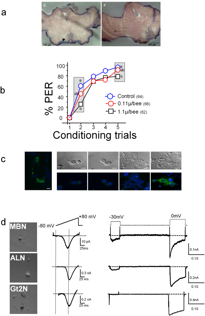
Figure 3. Neuronal CaV3 Ca2+ channel and odor learning.
(a) In situ hybridization of frontal section of honeybee brain using an Am-CaV3 specific RNA probe. Left, Am-CaV3 probe; right, control with the sens probe. The MBN stained by the probe is marked by an arrowhead. Scale bar 100 μm. (b) Standard PER conditioning assays performed on bees injected with 1.1 μg/bee mibefradil (black squares) or solvent (blue hexagons) depicted mild deficits in acquisition. At 0.11 μg/bee, mibefradil (red circles) had no effect. See Fig. S4a for tests on 1 h memory and olfactory generalization. (c) Staining using Am-CaV3 3CT6 antibody (at 1/100) of Hek-293 cells transfected with the Am-CaV3 or of various honeybee neurons in culture. Staining was performed 2 days after transfection, or at Div 2–6 for cultured neurons. LEFT. Staining of Am-CaV3-expressing HEK 293 cells display a cytoplasmatic and membrane localisation of the channel. RIGHT. Phase contrast images (top) and fluorescent images (bottom) of (from left to right) antennal lobe neurons (ALN), suboesophageal neurons (SBON), mushroom body neurons (MBN) and brain cells other than SBON, ALN and MBN. A few cells amongst the latter brain cells are stained by 3CT6 antibody. Scale bar: 10 μm. (d) Ba2+ currents recorded on MBN, ALN or Gt2N, during voltage ramps from −80 to +80 mV (middle) or during a two voltage-steps protocol from −100 mV to −30 mV and 0 mV (right). DIC (differential interference contrast) images of neurons where Ba2+ currents displayed on the left have been recorded. Scale bar, 10 μm.

