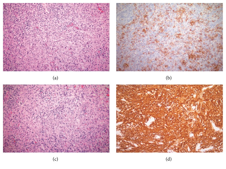
Figure 1.
(a) Both components of the histologic picture are seen in this section. Small-to-medium-sized lymphoid cells with clear cytoplasm and eosinophils are segregated by intervening whorls of spindly follicular dendritic cells (H&E ×320). (b) Numerous PD1+ cells are found in the cellular area of the lesion consistent with AITL (Immunohistochemistry with DAB ×320). (c) Several whorls of follicular dendritic cells are prominent in this section (H&E ×320). (d) The spindle follicular dendritic cells show very strong CD21 immunostaining. Note the negative staining related to the high endothelial venules (Immunohistochemistry with DAB ×320).

