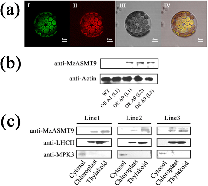
Figure 3. The localization of MzASMT9 protein in chloroplasts.
(a) The localization of MzASMT9-GFP in protoplasts of transgenic Arabidopsis ectopically expressing MzASMT9. It was visualized by confocal microscopy (60 × 10). The green fluorescence from the MzASMT9-GFP was shown in picture I. The chloroplasts were marked by the red fluorescence in picture II. The protoplast was observed under bright field in picture III. The green fluorescence and red fluorescence merged together in picture IV under bright field. (b) The specificity of anti-MzASMT9, as determined using Western blot of total protein from wild type and transgenic lines ectopically expressing MzASMT1 and MzASMT9. (c) The sub-organelle localization of MzASMT9-GFP in different transgenic Arabidopsis lines ectopically expressing MzASMT9 as determined by Western blot with antibodies of anti-MzASMT9, anti-LHCII or anti-MPK3. The anti-MPK3 antibody was used as a cytosolic marker; anti-LHCII was used as a thylakoids marker.

