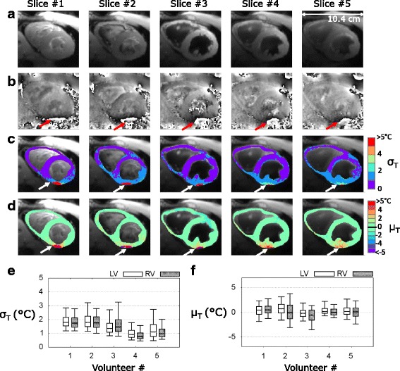
Fig. 2.

CMR thermometry evaluation on 5 healthy volunteers in short axis view. Typical results obtained on a volunteer (#5) are displayed: a averaged registered magnitude over 2 min 30 s of acquisition and (b) phase image presenting aliasing at the heart-liver-lung interface (red arrows). Temporal standard deviation σT (c) and mean μT (d) temperature maps are overlaid on averaged magnitude in a handmade ROI surrounding the all myocardium. White arrows indicate degraded CMR thermometry performance induced by remaining phase wraps. Pixels of this area with aberrant results (σT > 7 °C) were excluded from the statistical analysis presented in (e-f). Box-and-whisker plots of standard deviation σT (e) and mean μT (f) of the distribution of temperature in handmade ROIs surrounding the LV and the RV. For each graph, the levels of temperature correspond to 10% (lower horizontal bar), 25% (lower limit of the box), median value (horizontal line inside the box), 75% (upper limit of the box) and 90% (upper horizontal bar) of the distribution
