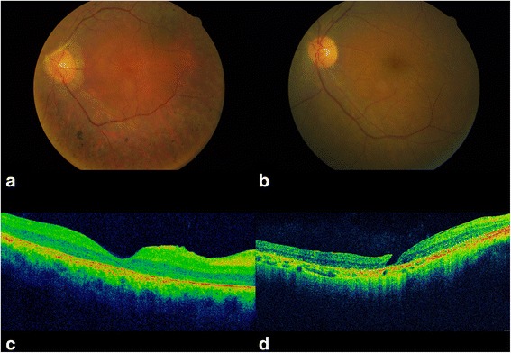
Fig. 2.

Representative images of the ocular findings in the BBS patients analyzed. Fundus photography showing a narrowing of retinal blood vessels, diffuse retinal pigment epithelial dystrophy with pigment clusters in mid-periphery; b narrowing of retinal blood vessels, widespread tapetoretinal degeneration and absence of pigment clusters. OCT scan showing c vitreomacular traction syndrome with retinal pigment epithelium dystrophy; (d) a macular lamellar hole with retinal pigment epithelium dystrophy
