Fig. 2.

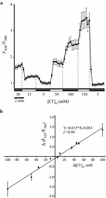
Calibration of the Cl-sensor in Neuro-2a cells. a Ratio for intensities of fluorescent signals emitted by Neuro-2a cells expressing Cl-sensor after excitation with 430 nm (F430) and 500 nm (F500) light. Neuro-2a cells were permeabilized with β-escin and incubated in extracellular solutions containing different Cl− concentrations [Cl−]o. An example of stepwise [Cl−]o changes is shown. Acquisition interval is 20 s. Mean values and the corresponding SEM are shown for 12 cells recorded in the optic field. b A linear regression for F430/F500 changes Δ(F430/F500) obtained for a broad range of [Cl−]o changes (Δ[Cl−]o) derived from multiple (n = 8) independent experiments similar to one shown in (a). Each data point shows how the F430/F500 ratio changes in response to a change of the extracellular chloride concentration Δ[Cl−]o. Error bars represent SEM. Error bars corresponding to −10, +5, and +10 mM data points are too small to be seen on the graph
