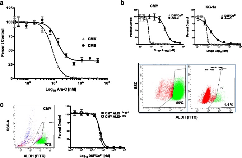
Fig. 1.

DSF/Cu2+ is cytotoxic in AML cell lines including Ara-C-resistant DS-AMKL cells independent of their endogenous ALDH activity. The DS-AMKL CMK and the non-DS-AMKL CMS cell lines were treated with different concentrations of Ara-C for 72 h and viability, expressed as percent of DMSO treated controls, was measured using CellTiter-Glo (CTG) assay. Drug dose response (DDR) curves show that DS-AMKL cell line CMK was more sensitive to Ara-C than the non DS-AMKL cell line CMS, with IC50 values of 71.16 nM and 170 nM, respectively (a). The DS-AMKL cell line CMY and the non-DS-AML cell line KG-1a were treated with different concentrations of Ara-C or DSF/Cu2+ for 72 h. CMY cells were relatively resistant to Ara-C (IC50 1131 nM), but sensitive to DSF/Cu2+ (IC50 83 nM). Although CMY cells had a 59% of ALDHbright cell population, and KG-1a cells had a 1.1% ALDHbright cell population (lower panel) they were both equally sensitive to DSF/Cu2+ (b). ALDH activity was measured using ALDELUORTM assay and flow cytometry. Dot plots in B show the percentage of ALDH-positive cells (FITC) on the x-axis, and the sideward scatter (SSC) on the y-axis. The gated cell populations were created using the ALDH inhibitor N, N-diethylaminobenzaldehyde (DEAB) provided with the kit. The ALDHbright and the ALDHlow cell populations from the CMY cell line were flow-sorted and treated with different concentrations of DSF/Cu2 for 72 h and the viability was measured using CTG (c). Viability is presented as percentage viable cell relative to control (DMSO-treated) (y-axis) plotted against the Log10 nM of drug concentrations. Experiments were repeated at least three times
