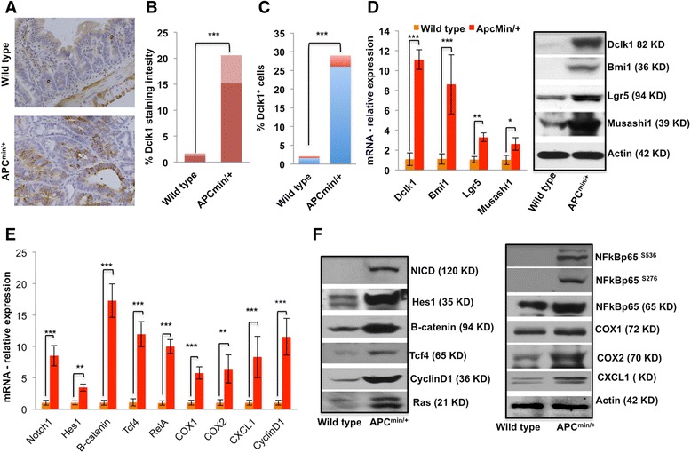
Fig. 2.

Increased expression of Dclk1 and Dclk1+ cells in the intestinal adenomas and adenocarcinomas of Apc Min/+ mice is associated with enhanced expression of tumor stem cell markers and pro-survival signaling. a IHC for Dclk1 in the small intestines of WT and Apc Min/+ mice. b Staining intensity was scored and is represented as a bar graph. c FACS data representing the % Dclk1+ cells isolated from the small intestines of WT and Apc Min/+ mice. d Differences in the number of Dclk1+ cells in staining and FACS corroborate with protein and mRNA levels of Dclk1 in the isolated IECs of WT and Apc Min/+ mice; protein and mRNA levels analyzed by western blot and RT-PCR of Bmi1, Lgr5, and Musashi1 in isolated IECs from WT and Apc Min/+ mice. f Protein expression levels of pro-survival signaling and their downstream targets in the isolated IECs of WT and Apc Min/+ mice, analyzed by western blot. e mRNA expression levels of pro-survival signaling and their downstream targets in the isolated IECs of WT and Apc Min/+ mice, analyzed by RT-PCR. All quantitative data are expressed as means ± SD of a minimum of three independent experiments. P values of <0.05 = *, <0.01 = **, and 0.001 = *** were considered statistically significant
