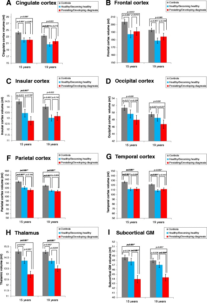
Fig. 2.

Brain volumetric differences between the two VLBW subgroups and controls at 15 and 19 years. The two VLBW diagnostic subgroups presented volume reductions in several cortices a-g and thalamus h compared with the control group. Subcortical GM reductions i were limited to the persisting/developing diagnosis VLBW subgroup. Results adjusted for age and sex. Subcortical structures adjusted for estimated intracranial volume. Abbreviations: GM: Gray matter. * Significant results after adjusting for multiple testing
