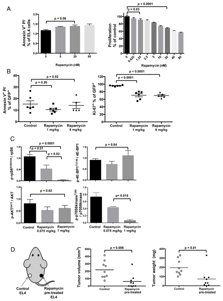
Figure 3. Rapamycin directly inhibits EL4 mTORC1 signals and proliferation.
(A) EL4 cells were treated with rapamycin or vehicle control for 72 h in vitro and stained with propidium iodide (PI) and Annexin V to evaluate apoptosis by flow cytometry. MTT assay was used to assess proliferation. n=6/group. P values, t-test. (B) Apoptosis and Ki67 (proliferation marker) of ex vivo tumor cells from Rag2 KO mice after rapamycin treatment (1 or 8 mg/kg) for 16 days, stained and analyzed by flow cytometry. P values, one-way ANOVA. (C) EL4-challenged WT mice were treated with rapamycin (0.075 or 1 mg/kg) or vehicle control daily from day 4 and sacrificed on day 21 with composite quantification shown from 3 Western blots of tumor lysates. P values, t-test. n=4/group for each blot. Error bars, mean ± SEM. (D) Tumor volumes and weights of EL4 cells pre-treated with rapamycin 100 nM or PBS for 48 h in vitro and then injected (40,000 cells) s.c. into WT mice (1 treated and 1 untreated tumor challenged in one mouse, on opposite flanks, as depicted on the left). N= 10/group. Tumors were measured with calipers and resected for weight one week later. P values, unpaired t test.

