
Figure 4.
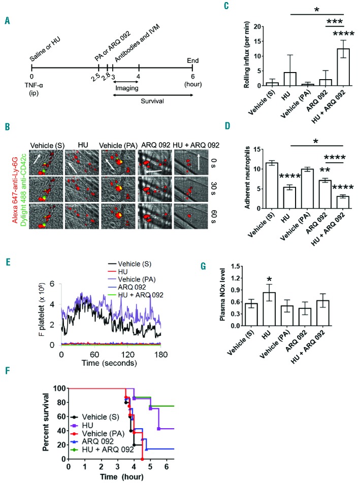
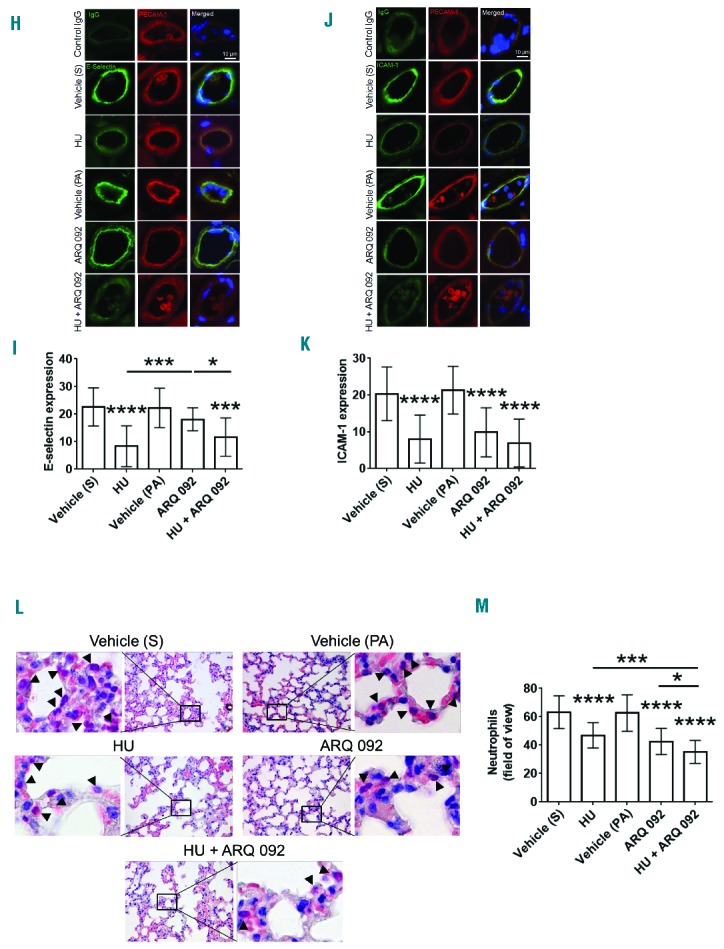
(A–G). Oral administration of hydroxyurea and ARQ 092 has numerous beneficial effects in TNF-α-challenged SCD mice. Intravital microscopy was performed as described in the Methods. Neutrophils and platelets were labeled by infusion of Alexa Fluor 647-conjugated anti-Ly-6G and DyLight 488-conjugated anti-CD42c antibodies, respectively. (A) Timeline for each treatment and surgery for intravital microscopy (IVM) in SCD mice. S: saline; HU: hydroxyurea; PA: 0.01 M phosphoric acid. (B) Representative images at various time-points. The time “0” was set as the image capture was initiated for each vessel. (C, D) Number of rolling (for 1 min) and adherent neutrophils (for 5 min). Data represent the mean ± SEM (n = 7–8 mice). (E) The integrated median fluorescence intensities of anti-CD42c antibodies (F platelets) were plotted over time. (F) Survival curves. Survival was significantly improved in the groups treated with hydroxyurea alone (P = 0.0028) or both hydroxyurea and ARQ 092 (P=0.0017), compared to each vehicle control. Mantel-Cox log-rank test. (G) Plasma NOx levels were measured as described in the Methods. Data represent the mean ± SD (n = 7–8 mice per group). (H–M). Oral administration of hydroxyurea and ARQ 092 has numerous beneficial effects in TNF-α-challenged SCD mice. (H–K) After recording survival times, the cremaster muscle was taken out for immunohistochemistry. Muscle sections were labeled with control IgG or rat monoclonal antibodies against E-selectin or ICAM-1 and then with DyLight 488-conjugated anti-rat IgG antibodies, followed by incubation with APC-conjugated anti-PECAM-1 antibodies and a mounting reagent containing DAPI. (H and J) Representative images. Bar = 10 μm. (I and K) The fluorescence intensities of antibodies. Data represent the mean ± SD (n = 18–22 vessels in 5 mice per group). (L–M) After recording survival times, lungs were taken out for histochemistry. Neutrophils were stained with naphthol AS-D chloroacetate. (L) Representative images. (M) The number of transmigrated neutrophils (arrow heads) was quantified in the field of view (110 mm2). Data represent the mean ± SD (n = 36–42 sections in 5–6 mice per group). *P<0.05, **P<0.01, ***P<0.001, and ****P<0.0001 versus each vehicle control (or between two groups), ANOVA and the Tukey test. S: saline; HU: hydroxyurea; PA: phosphoric acid.