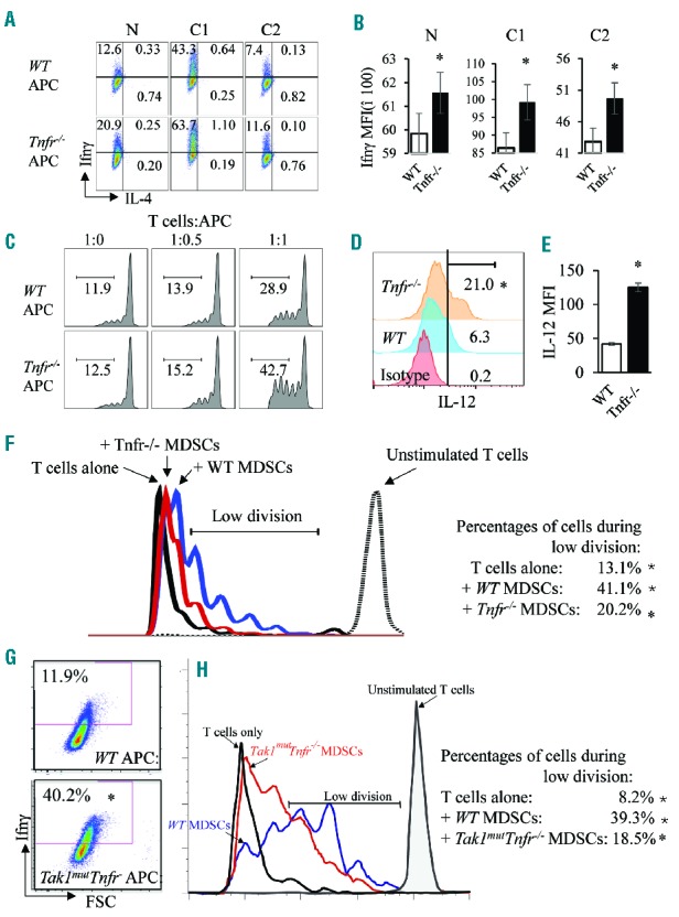
Figure 4.

Enhanced function of Tnfr−/− APCs and decreased function of Tnfr−/− MDSCs. (A and B) T cells from wild-type (WT) spleens were first isolated using a pan-T-cell isolation kit, then separated with anti-CD62L. Naïve T cells (CD62L+) were further cultured with WT and Tnfr−/− APCs for seven days under three conditions: 1) neutral condition (N); 2) Th1-priming condition (C1, in the presence of IL-12 and anti-IL-4); and 3) Th2-priming condition (C2, in the presence of IL-4 and anti-IL-12). Cells were collected and subjected to intracellular staining for cytokines (A). The mean fluorescence intensity (MFI) of Ifnγ in all of the groups was calculated and is presented in bar graphs (B). (C–E) CFSE-labeled WT T cells were co-cultured with either WT or Tnfr−/− APCs in the presence of anti-CD3 and anti-CD28. After three days of culturing, cells were collected and analyzed for the percentage of T cells with more than 3 divisions (C). WT and Tnfr−/− APCs were stained for intracellular IL-12 (D). MFI of IL-12 in the two groups in (D) was calculated and presented in bar graphs (E). (F) CD11b+Gr1+ cells were sorted from WT and Tnfr−/− bone marrow and added to anti-CD3-activated, CFSE-labeled WT T cells at a ratio of T:MDSC=1:2 and co-cultured for six days. The CFSE signals were analyzed on day 6. The results were analyzed to show the cells with a low number of divisions (2–6 divisions). (G) T cells from WT spleens were isolated using a pan-T-cell isolation kit and were cultured with WT and Tak1mutTnfr−/− APCs for seven days under Th1-priming condition. Cells were collected and subjected to intracellular staining for Ifnγ. (I) CD11b+Gr1+ cells were sorted from WT and Tak1mutTnfr−/− BM, added to anti-CD3-activated, CFSE-labeled WT T cells at a ratio of T:MDSC=1:2 and co-cultured for six days. The CFSE signals were analyzed on day 6. The results were analyzed to show the cells with a low number of divisions. The samples were analyzed in triplicate and experiments were repeated independently twice. *P<0.05 compared to WT.
