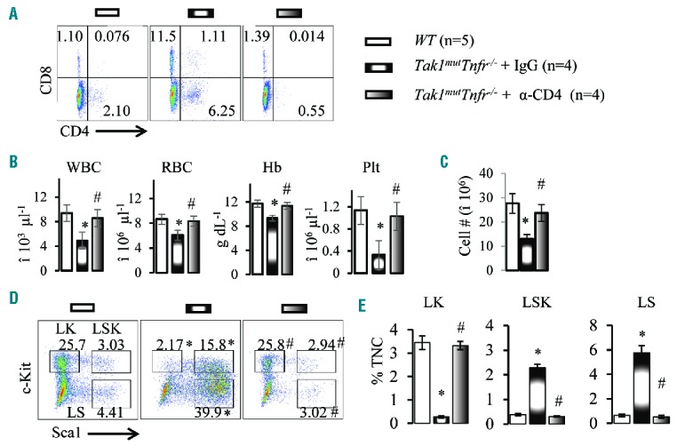
Figure 5.

Depletion of CD4+ T cells restores normal hematopoiesis to Tak1mutTnfr−/− mice. (A) Bone marrow (BM) samples were collected from wild-type (WT) mice and Tak1mutTnfr−/− mice treated with IgG and anti-CD4 antibody. Flow cytometric analyses of the frequencies of CD4+ and CD8+ T cells are shown. (B) White blood cells (WBC), red blood cells (RBC), hemoglobin (Hb) and platelets (plt) were analyzed using the Hemavet 950 Hematology System. (C) Number of total nucleated cells (TNCs) in BM from two hind limbs were counted and compared. (D) Representative flow cytometric plots for analysis of BM hematopoietic stem cells (HSCs) and hematopoietic progenitor cells (HPCs). BM cells were first gated on the Lin− population and then analyzed for LK, LSK and LS populations. (E) Percentages of LK, LSK and LS populations in TNCs of BM. Data are presented as means±SD. *P<0.05 compared to WT; #P<0.05 compared to IgG treatment.
