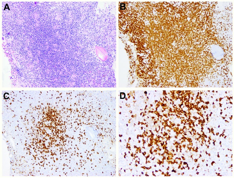
Figure 2.

Nodular lymphoid infiltrates containing mixed T and B cells in a bone marrow with ALPS (case l8). (A) H&E, X 40, (B) CD3, X 40, (C) CD45RO, X 40, (D) CD20, X 200. Note the clusters of medium-sized to large B cells surrounded by numerous small T cells. There is loss of expression of CD45RO.
