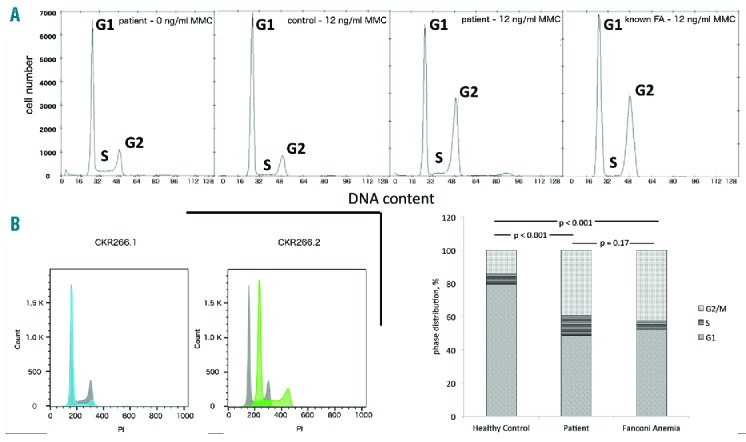
Figure 2.

Disturbance of DNA repair in patient samples. (A) Mitomycin C (MMC) sensitivity assay - patient fibroblasts (without MMC, left chart) were exposed to MMC (12 ng/ml) and cultured for 48h. Staining with 4′,6-diamidino-2-phenylindole (DAPI) shows an increased number of G2 phase cells, indicating that cells are in a cycle arrest after induced damage by MMC, a known DNA intercalating agent (second right chart). A G2 increase is not seen in healthy controls (second left chart), but detected in a similar quantity in an internal control with fibroblasts from a MMC sensitive patient control (right chart). Cell cycle distribution among mentioned samples – a χ2 test shows a significant difference between healthy control and patient sample and MMC sensitive patient control, while there is no difference between patient sample and MMC sensitive patient control (bottom right chart) (B) Flow cytometric DNA content measurement of two different LCLs generated from patient’s bone marrow stained with PI following ethanol fixation. Both cell lines were cultured and processed under the same standard conditions from the same specimen. Left: Patient LCL#1 cells (blue) show a slightly increased DNA amount than normal control LCLs (grey). Right: Patient LCL#2 cells (green) show aneuploidy (polyploidy in another experiment), compared to healthy human samples. PI: propidium iodide; FA: fanconi anemia; MMC: mitomycin C.
