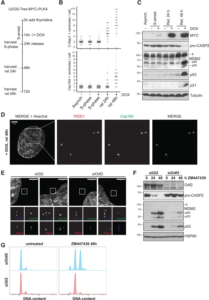
Figure 6.
Extra mother centrioles activate the PIDDosome. (A) Scheme of the protocol used to time-resolve the appearance of extra centrioles at different maturation stages and PIDDosome activation. U2OS-Trex-MYC-PLK4 cells were arrested in S phase with thymidine. After 14 h, they were either left untreated or induced for an additional 10 h with doxycyline (Dox). Cells were either processed for immunofluorescence (Supplemental Fig. S10A) or immunoblot directly during S arrest or released for 24 and 48 h. (B) Scatter plot for the abundance of C-Nap1-positive and Cep164-positive centrioles assessed in 50 individual cells. (C) PIDDosome activation was followed by immunoblot analysis using the indicated antibodies. (D) Localization of PIDD1 at extra mother centrioles generated by PLK4 overexpression. (E–G) A549 cells transfected with the indicated siRNAs were subjected to immunofluorescence with the indicated antibodies (E) or treated with ZM447439 for the indicated times and subjected to either immunoblotting (F) or DNA content analysis (G).

