. 2016 Dec 23;47(1):123–131. doi: 10.4070/kcj.2016.0203

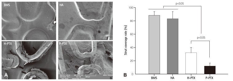
Fig. 7. The strut coverage rate. (A) Representative SEM images of the luminal area of the stents. The latter were harvested at 4 weeks postimplantation. They were bisected longitudinally and subjected to SEM examination. (B) Histomorphometric analysis of strut coverage rate ntified by ntified by special software (Visus 2000 Visual Image Analysis System; IMT Tech., San Diego, CA, USA). (Visus 2000 Visual Image Analysis System; IMT Tech., San Diego, CA, USA). special software. BMS: bare-metal stent, HA: hyaluronic acid, H-PTX: hyaluronic-acid-coated paclitaxel-eluting stent, P-PTX: poly(L-lactide)-coated paclitaxel-eluting stent, SEM: scanning electron microscope.

Fig. 7