Fig. 4.
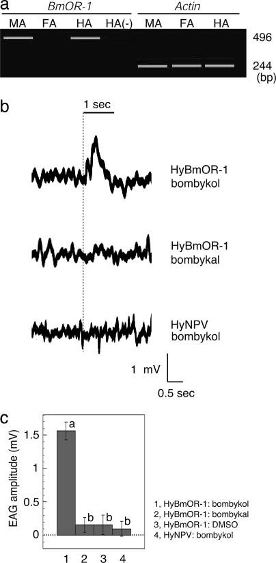
EAG responses of the HyBmOR-1-infected female adult antennae to bombykol. (a) Ectopic expression of BmOR-1 in day-0 female adult antennae infected with BmOR-1 recombinant baculovirus (HyBmOR-1) at 4 days before eclosion. RT-PCR products by using RNA isolated from the antennae of the moths were separated by electrophoresis. MA, male antennae; FA, female antennae; HA, HyBmOR-1-infected female antennae. Minus sign indicates RT-PCR was performed without reverse transcriptase. (b) EAG recordings of HyBmOR-1- or HyNPV-infected female antennae exposed to bombykol or bombykal at 500 ml/min. EAG traces of the same HyBmOR-1-infected female antenna to bombykol (Top) or bombykal (Middle) are shown. Stimulus was applied starting at the time indicated by the dotted line for the duration of 1 s, as indicated by the solid line above the recording profiles. In these recordings, a reference electrode was placed at the tip of antenna, and thereby, the response was shown as a positive signal. (c) Quantitative analysis of EAG responses of HyBmOR-1-infected female antennae to bombykol. Amplitudes of responses of HyBmOR-1-infected female antennae to bombykol (lane 1) or bombykal (lane 2) (4 μg on filter paper) or to 1% DMSO as a control (lane 3) with a flow rate of 350 ml/min (n = 10 each) were quantified. EAG recordings to these three stimuli were measured on the same antenna. WT HyNPV was used as a negative control (lane 4) (n = 25). Error bars represent ± SEM. Average values marked by different letters are significantly different (Scheffé's test: P < 0.05). The EAG response of HyBmOR-1 to bombykol (lane 1) was significantly larger than to bombykal (lane 2) or DMSO (lane 3). The other three responses (lanes 2–4) were not significantly different.

