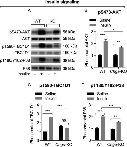
Figure 2.
Assessment of insulin-induced signaling by Western blot in GAS muscle. Mice were treated with saline or insulin (0.4 U/g BW) for 10 min before tissue harvesting. (A) Western blots showing the expression of phospho-AKT at Ser473, total AKT, phospho-TBC1D1 at Thr590, total PBC1D1, phospho-P38 at Thr180/Tyr182 and total P38 in WT and Chga-KO mice. Densitometric values showing pS473-AKT/total AKT (B), pT590-TBC1D1/total TBC1D1 (C), and pT180/Y182-P38/total P38 in WT and Chga-KO mice after endurance exercise. Note compromised insulin-induced signaling in Chga-KO mice.

