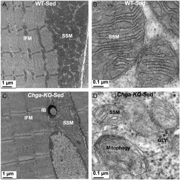
Figure 8.
Subsarcolemmal mitochondria (SSM) in sedentary WT and Chga-KO GAS muscle. (A) SSM and IFM at magnification 2500× in WT mice. (B) SSM at magnification 30,000× in WT mice. Note cristae emerging from the inner mitochondrial membrane. (C) SSM and IFM at magnification 2500× in Chga-KO mice. Note inclusion body in the subsarcolemmal region. (D) SSM at magnification 30,000× in Chga-KO mice. Note mitophagy, fewer cristae and glycogen granules (GLY).

