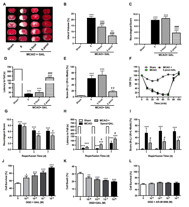
Figure 2.

Galanin treatment protects neurons against MCAO and OGD-induced ischemic injuries. (A and B) Representative TTC staining and graphs, showing significant decrease of infarct volume of ischemic stroke mice after 5 fmol and 5 pmol GAL pre-treatment (n=6 per group); (C) Neurological scores of mice after 1 h MCAO and 24 h reperfusion showed a significant attenuation of deficits at 5 pmol (but not 5 fmol) GAL pre-treatment (n=8 per group); (D) Evaluation of muscle strength using the wire hanging test. All sham group mice could stay on the wire mesh for the maximum observation period of 600s, but MCAO mice showed a significant decrease of latency time. 5 pmol GAL pre-treatment also showed a significant improvement, but still significantly lower than the sham group (n=8 per group); (E) Evaluation of limb coordination via the cylinder test shows a marked increase of contralateral limb usage in mice receiving 5 pmol GAL pre-treatment (n=8 per group); (F) Cerebral blood flow (CBF) of the middle cerebral artery detected via Laser Doppler flowmetry, which shows a significant decrease of CBF after insertion of the intraluminal monofilament, but no changes were observed after the i.c.v injection of GAL (n=5 per group); (G) Neurological scores of mice after 1 hr MCAO/3-7 d reperfusion showed a significant attenuation of deficits after 5 pmol GAL post-treatment (n=8 per group); (H) Evaluation of muscle strength via the wire hanging test. MCAO mice showed a significant decrease of latency time. Conversely, the 5 pmol GAL post-treatment group showed a significant improvement (n=8 per group); (I) Evaluation of limb coordination the cylinder test, showing a marked increase of contralateral limb usage of ischemic stroke mice receiving 5 pmol GAL after reperfusion (n=8 per group); (J) The MTS assay results show that pre-incubation with GAL 15 min prior to OGD could increase neuronal survival rates in a dosage dependent manner in primary cultured cortical neurons after 1 h OGD/24 hr reoxygenation (n=6 per group); (K) The cytotoxicity assay results indicate a significant decrease of cell death rates in GAL-treated group when compared with OGD group (n=6 per group); (L) MTS assay demonstrate that pre-incubation with the GalR2/3 agonist AR-M1896 at various concentrations 15 min prior to OGD did not affect neuronal survival rates of primary cultured cortical neurons after 1 h OGD/24 h reoxygenation (n=6 per group). Data are presented as mean ± SEM; *p<0.05, **p<0.01, ***p<0.001 vs. sham/0 M GAL; #p<0.05, ##p<0.01, ###p<0.001 vs. MCAO group.
