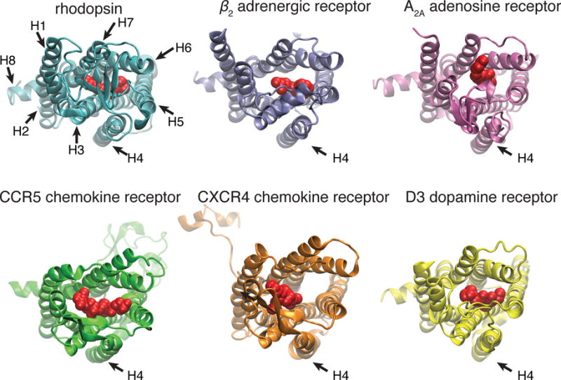
Figure 1.

Comparison of the crystal structures of different Class A GPCRs with bound ligands: 1) rhodopsin and 11-cis-retinal (protein data base access code 1GZM);[46] 2) β2 adrenergic receptor and carazolol (2RH1);[47] 3)A2A adenosine receptor and ZM241385 (3EML);[48] 4) CCR5 chemokine receptor and maraviroc (4MBS);[49] 5) CXCR4 chemokine receptor and IT1t (3ODU);[50] 6) D3 dopamine receptor (3PBL),[51] viewed from the EC side, with the ligands shown in red. The TM4 of these receptors (indicated by an arrow) are positioned outside the helix bundle and not in contact with the ligands.
