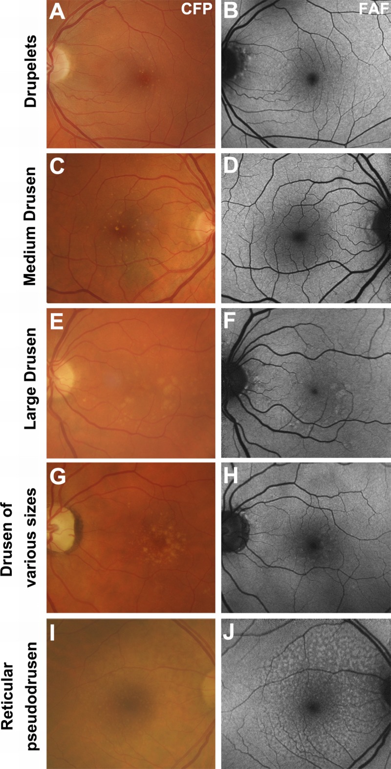
FIGURE 2.

The appearance of drusen subtypes using fundus autofluorescence. All images are presented in pairs. A–B, Drupelets (small drusen) and (C–D) medium-sized drusen causing a negligible effect on the fundus autofluorescence image. E–F, Large, soft indistinct drusen, which appear as well-defined patches of hyper-autofluorescence. G–H, Drusen varying in size from small to large, showing their variable effects. I–J, Reticular pseudodrusen, appearing as multiple, clustered, regularly networked, round areas of low-contrast hypo-autofluorescence. Abbreviations as in Fig. 1.
