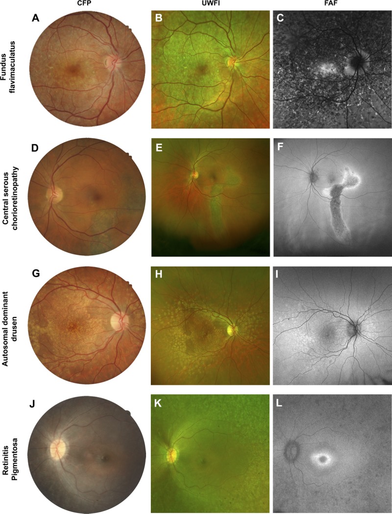
FIGURE 6.

Case series from patients that featured more extensive fundus changes involving the macula. A single case is displayed across the row. A–C, Fundus flavimaculatus: the characteristic flecks displayed prominent hyper-autofluorescence. D–F, Central serous chorioretinopathy distinguished by the typical appearance of a gravitational atrophic tract. G–I, Autosomal dominant drusen which appeared as discrete, radially distributed areas of hyper-autofluorescence. J–L, Retinitis pigmentosa characterized by patchy hypofluorescence in the periphery and a hyper-autofluorescent ring at the fovea. Abbreviations as in Fig. 1.
